Publications
Preprints
Multi-stage efficient coding of perception and value in goal-directed behavior ()
Saurabh Bedi, Gilles de Hollander, Maximilian V. Harl, Christian C. Ruff
bioRxiv 2026.06.08.730978 doi: 10.64898/2026.06.08.730978
A Neural Population Code for Value in Human Orbitofrontal Cortex ()
Raphaël Le Bouc, Gilles de Hollander, Marcus Grueschow, Shira M. Lupkin, Vincent B. McGinty, Rafael Polania, Christian C. Ruff
bioRxiv 2026.03.28.715037 doi: 10.64898/2026.03.28.715037
Separable neurocomputational mechanisms underlying multisensory learning ()
Saurabh Bedi, Ella Casimiro, Gilles de Hollander, Nina Raduner, Fritjof Helmchen, Silvia Brem, Arkady Konovalov, Christian C. Ruff
bioRxiv 2025.11.18.688925 doi: 10.1101/2025.11.18.688925
Acute stress reduces risk-aversion by changing magnitude perception ()
Maike F. Renkert*, Gilles de Hollander*, Gökhan Aydogan, Saurabh Bedi, Paul Forbes, Claus Lamm, Christian C. Ruff
bioRxiv 2025.11.03.685504 doi: 10.1101/2025.11.03.685504
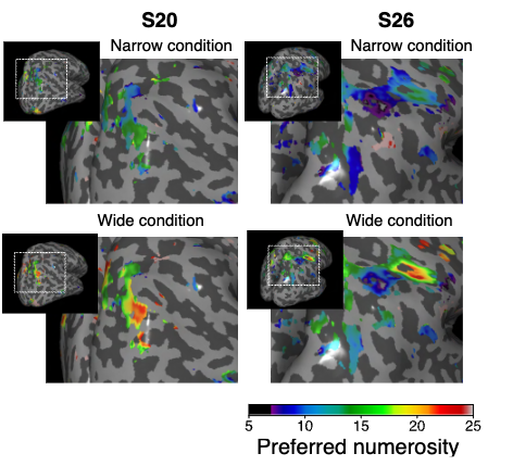
Distributed range adaptation in human parietal encoding of numbers ()
Arthur Prat-Carrabin*, Gilles de Hollander*, Saurabh Bedi, Samuel J. Gershman, Christian C. Ruff
bioRxiv 2025.09.25.675916 doi: 10.1101/2025.09.25.675916
Probability weighting arises from boundary repulsions of cognitive noise ()
Saurabh Bedi, Gilles de Hollander, Christian C. Ruff
bioRxiv 2025.09.11.675565 doi: 10.1101/2025.09.11.675565
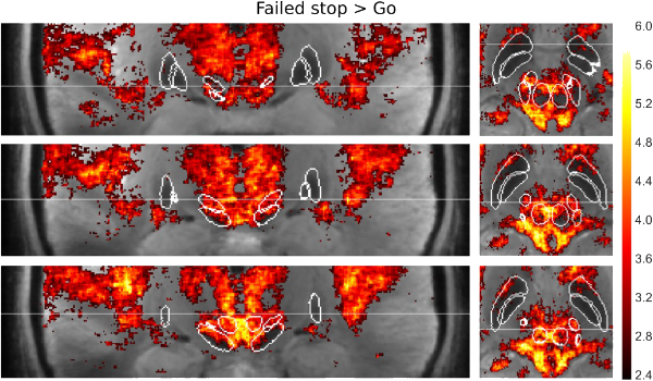
Risk preferences causally rely on parietal magnitude representations: Evidence from combined TMS-fMRI ()
Gilles de Hollander, Marius Moisa, Christian C. Ruff
bioRxiv 2025.01.13.632678 doi: 10.1101/2025.01.13.632678
Rapid Changes in Risk Preferences Originate from Bayesian Inference on Parietal Magnitude Representations ()
Gilles de Hollander, Marcus Grueschow, Franciszek Hennel, Christian C. Ruff
bioRxiv 2024.08.23.609296 doi: 10.1101/2024.08.23.609296
A retinotopic reference frame for space throughout human visual cortex ()
Martin Szinte*, Gilles de Hollander*, Marco Aqil*, Ines Verissimo, Serge Dumoulin, Tomas Knapen
bioRxiv 2024.02.05.578862 doi: 10.1101/2024.02.05.578862
2025
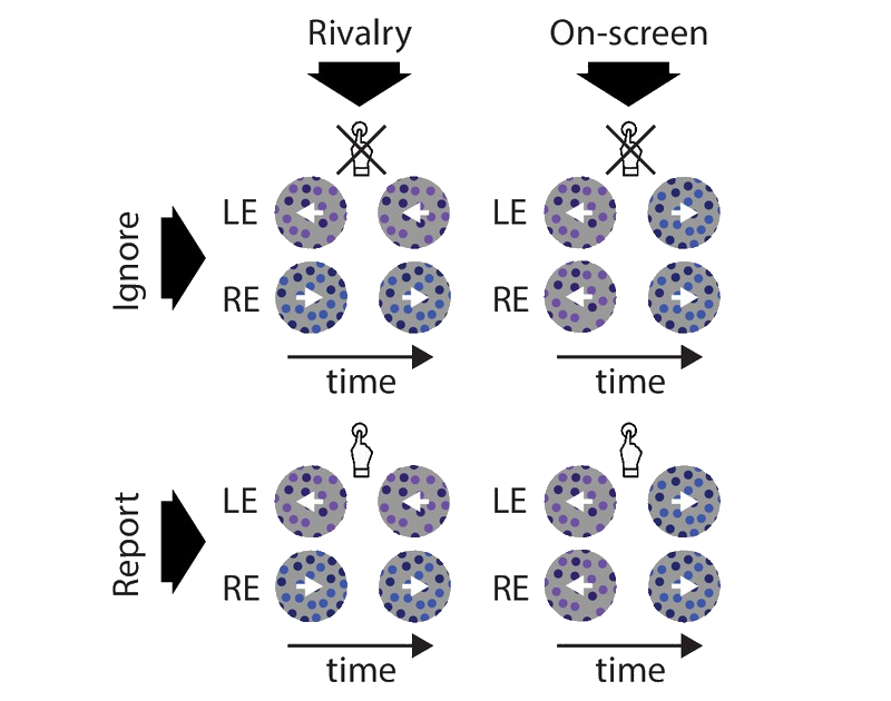
Mesoscale cortical mechanisms of perceptual conflict resolution in binocular rivalry ()
Chencan Qian, Zihao Zhang, Zhiqiang Chen, Gilles de Hollander, Tomas Knapen, Sheng He, Peng Zhang
Nature Human Behaviour Qian2025 doi: 10.1038/s41562-025-02320-4
Psilocybin alters visual contextual computations ()
Marco Aqil, Gilles de Hollander, Nina Vreugdenhil, Tomas Knapen, Serge O. Dumoulin
Nature Communications 16:10271 doi: 10.1038/s41467-025-65150-y
2024
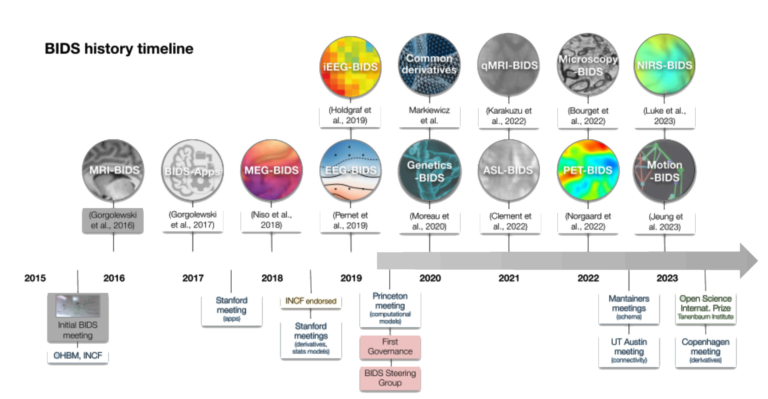
The Past, Present, and Future of the Brain Imaging Data Structure (BIDS) ()
Russell A. Poldrack, Christopher J. Markiewicz, Stephan Appelhof, ..., Gilles de Hollander, ..., Krzysztof J. Gorgolewski
Imaging Neuroscience 2309.05768 doi: 10.1162/imag_a_00103
No difference in prior representations of what to attend and what to ignore ()
Michel Failing, Gilles de Hollander, Stefan Pollmann, Christian N. L. Olivers
Journal of Cognitive Neuroscience doi: https://doi.org/10.1080/13506285.2024.2338605
2023
Individual risk attitudes arise from noise in neurocognitive magnitude representations. ()
Miguel Barretto García*, Gilles de Hollander*, Grueschow, M., Rafael Polanía, Michael Woodford, Christian C. Ruff
Nature Human Behavior 2022.08.22.504413 doi: 10.1038/s41562-023-01643-4
2022
7T functional MRI finds no evidence for distinct functional subregions in the subthalamic nucleus during a speeded decision-making task ()
Steven Miletić, Max C. Keuken, Martijn Mulder, Robert Trampel, Gilles de Hollander*, Birte U. Forstmann*
Cortex j.cortex.2022.06.014 doi: 10.1016/j.cortex.2022.06.014
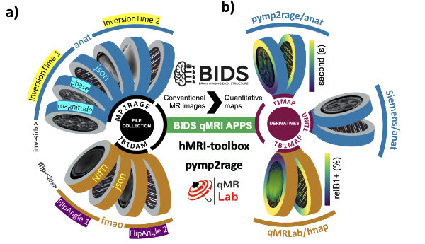
qMRI-BIDS: an extension to the brain imaging data structure for quantitative magnetic resonance imaging data ()
Agah Karakuzu, Stefan Appelhoff, Tibor Auer, Mathieu Boudreau, Franklin Feingold, Ali R Khan, Alberto Lazari, Christopher J Markiewicz, Martijn J Mulder, Christophe Phillips, Taylor Salo, Nikola Stikov, Kirstie Whitaker*, Gilles de Hollander*
Nature Scientific Data 2021.10.22.21265382 doi: 10.1038/s41597-022-01571-4
2021
Separable pupillary signatures of perception and action during perceptual multistability ()
Jan W Brascamp, Gilles de Hollander, Michael D Wertheimer, Ashley N DePew, Tomas Knapen
eLife 10:e66161 doi: 10.7554/eLife.66161
Ultra-high field fMRI reveals origins of feedforward and feedback activity within laminae of human ocular dominance columns ()
Gilles de Hollander, Wietske van der Zwaag, Chencan Qian, Peng Zhang, Tomas Knapen
Neuroimage 228, 117683 doi: 10.1016/j.neuroimage.2020.117683
2019
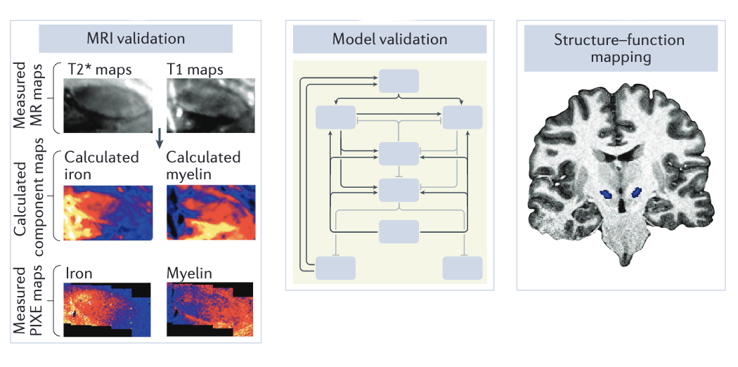
MP2RAGEME: T1, T2*, and QSM mapping in one sequence at 7 tesla ()
Matthan WA Caan, Pierre-Louis Bazin, José P Marques, Gilles de Hollander, Serge O Dumoulin, Wietske van der Zwaag
Human Brain Mapping 40(6), 1786– 1798 doi: 10.1002/hbm.24490
A neural substrate of early response capture during conflict tasks in sensory areas ()
Yael Salzer, Gilles de Hollander, Leendert van Maanen, Birte U Forstmann
Neuropsychologia 124, 226-235 doi: 10.1016/j.neuropsychologia.2018.12.009
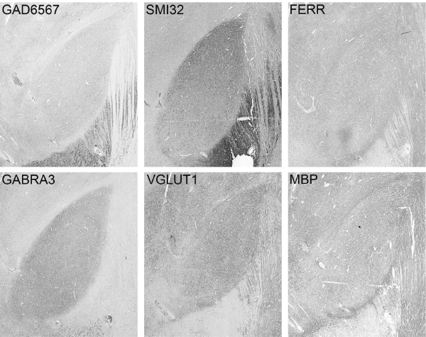
The Functional Microscopic Neuroanatomy of the Human Subthalamic Nucleus ()
Anneke Alkemade*, Gilles de Hollander*, Steven Miletic*, Max C Keuken, Rawien Balesar, Onno de Boer, Dick F Swaab, Birte U Forstmann
Brain Structure and Function 224, 3213–3227 doi: 10.1007/s00429-019-01960-3
The Importance of Standards for Sharing of Computational Models and Data ()
Russell A Poldrack, Franklin Feingold, Michael J Frank, Padraig Gleeson, Gilles de Hollander, Quentin J M Huys, Bradley C Love, Christopher J Markiewicz, Rosalyn Moran, Petra Ritter, Timothy T Rogers, Brandon M Turner, Tal Yarkoni, Ming Zhan & Jonathan D Cohen
Computational Brain and Behavior 2, 229-232 doi: 10.1007/s42113-019-00062-x
2017
Towards a mechanistic understanding of the human subcortex ()
Birte U Forstmann, Gilles de Hollander, Leendert van Maanen, Anneke Alkemade, Max C Keuken
Nature Reviews Neuroscience 18, 57–65 doi: 10.1038/nrn.2016.163
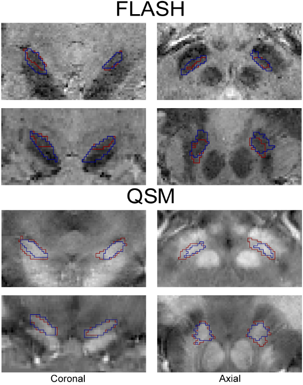
Comparing functional MRI protocols for small, iron-rich basal ganglia nuclei such as the subthalamic nucleus at 7 T and 3 T ()
Gilles de Hollander, Max C Keuken, Wietske van der Zwaag, Birte U Forstmann, Robert Trampel
Human Brain Mapping 38:3226–3248 doi: 10.1002/hbm.23586
Comparison of T2*- weighted and QSM contrasts in Parkinson's disease to visualize the STN with MRI ()
Anneke Alkemade*, Gilles de Hollander*, Max C Keuken, Andreas Schäfer, Derek VM Ott, Johannes Schwarz, David Weise, Sonja A Kotz, Birte U Forstmann
PLOS ONE 12(4), e0176130 doi: 10.1371/journal.pone.0176130
Sensory neural pathways revisited to unravel the temporal dynamics of the Simon effect: A model-based cognitive neuroscience approach. ()
Yael Salzer, Gilles de Hollander, Birte U Forstmann
Neuroscience & Biobehavioral Reviews 77, 48-57 doi: 10.1016/j.neubiorev.2017.02.023
2016
The Age-ility Project (Phase 1): Structural and functional imaging and electrophysiological data repository. ()
Frini Karayanidis, Max C Keuken, Aaron Wong, Jaime L Rennie, Gilles de Hollander, Patrick S Cooper, W Ross Fulham, Rhoshel Lenroot, Mark Parsons, Natalie Phillips, Patricia T Michie, Birte U Forstmann
Journal of Neuroscience 124(b), 1137-1142 doi: 10.1016/j.neuroimage.2015.04.047
Combining Computational Models of Cognition and Neural Data to Learn about Mixed Task Strategies ()
Gilles de Hollander
Journal of Neuroscience 36(1), 1-3 doi: 10.1523/jneurosci.3690-15.2016
Different Ways of Linking Behavioral and Neural Data via Computational Cognitive Models ()
Gilles de Hollander, Scott SD Brown, Birte U Forstmann
Biological Psychiatry: Cognitive Neuroscience and Neuroimaging 1 (2): 101-109 doi: 10.1016/j.bpsc.2015.11.004
Transcranial direct current stimulation does not influence the speed-accuracy tradeoff in perceptual decision-making: Evidence from three independent studies. ()
Gilles de Hollander, Ludovica Labruna, Roberta Sellaro, Anne Trutti, Lorenza S. Colzato, Roger Ratcliff, Richard B Ivry, Birte U Forstmann
Journal of Cognitive Neuroscience 28 (9): 1283-1294 doi: 10.1162/jocn_a_00967
2015
The subcortical cocktail problem; Mixed signals from the subthalamic nucleus and substantia nigra ()
Gilles de Hollander*, Max C Keuken*, Birte U Forstmann
PLOS ONE 10(3), e0120572 doi: 10.1371/journal.pone.0120572
2014
A gradual increase of iron toward the medial-inferior tip of the subthalamic nucleus ()
Gilles de Hollander*, Max C Keuken*, Pierre-Louis Bazin, Marcel Weiss, Jane Neumann, Katja Reimann, Miriam Wähnert, Robert Turner, Birte U Forstmann, Andreas Schäfer
Human Brain Mapping 35:4440–4449 doi: 10.1002/hbm.22485
2011
Summarization of meetings using word clouds ()
Gilles de Hollander, Maarten Marx
2011 CSI International Symposium on Computer Science and Software Engineering (CSSE) 12137845 doi: 10.1109/CSICSSE.2011.5963995